A radiation plan is only as useful as its match to the tumour, nearby organs and daily treatment conditions. By the end, you will understand what 3DCRT can improve, when another technique may offer a better balance, and which technical and clinical questions to ask before starting treatment.
Key takeaways
- 3DCRT shapes radiation beams around a tumour using planning CT images.
- Ask how 3DCRT protects nearby organs at the treated cancer site.
- Compare 3DCRT with IMRT, VMAT and stereotactic treatment before deciding.
- Bring your pathology report, scans and medication list to the planning visit.
How CT-Based 3DCRT Targets a Tumour
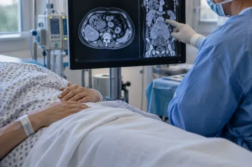
A planning CT scan gives the treatment team a three-dimensional map of the tumour and nearby normal tissue. The oncologist outlines these structures on each CT slice, then calculates radiation from several beam directions. Shaped beam apertures conform to the tumour’s outline, delivering the prescribed dose while limiting exposure beyond its edges.
Older two-dimensional radiotherapy relied mainly on plain X-rays and bony landmarks to aim rectangular fields. That approach could not show the tumour and organs at risk as precisely in three dimensions. CT-based 3DCRT can therefore produce a more accurate dose distribution and reduce unnecessary radiation to nearby normal tissue.
The benefit depends on the tumour’s position, size and movement. A tumour that shifts with breathing needs motion assessment through methods such as four-dimensional CT, breath-hold or image guidance; a static plan can miss the intended target.
When comparing 3D conformal radiation therapy in Pune, ask whether the centre documents treatment verification and a physics quality check. 3DCRT is not automatically the safest option: IMRT can create more complex dose patterns when a tumour wraps around a sensitive organ.
The Patient Benefits—and the Limits—Across Cancer Sites
3DCRT may benefit cancers in the prostate, breast, lung, head and neck, brain, pelvis and gastrointestinal tract when the tumour’s position and shape allow beams to cover it while limiting dose to nearby organs. It can aim for cure, reduce recurrence after surgery, support other treatment, or relieve pain, bleeding, obstruction or pressure.
| Cancer site | Possible advantage | Important limit |
|---|---|---|
| Prostate | Shapes dose around the prostate while limiting exposure to the rectum and bladder | Urinary, bowel and sexual side effects can still occur |
| Breast | Treats the breast or chest wall while reducing dose to the heart and opposite breast | Heart or lung exposure remains a concern, especially on the left side |
| Lung | Targets a lung tumour while limiting dose to healthy lung and spinal cord | Breathing motion can shift the target; four-dimensional CT, breath-hold or gating may be needed |
| Head and neck | Helps spare the spinal cord, brainstem, salivary glands and swallowing structures | Dry mouth, swallowing problems and skin reactions remain possible |
| Pelvic or gastrointestinal | Helps protect bowel, bladder, kidneys and spinal cord | Large or moving targets may require another planning technique |
The central radiation therapy benefits Pimpri Chinchwad patients seek are better anatomical targeting and deliberate organ sparing, not freedom from side effects. Skin irritation, tiredness, nausea, pain flare or site-specific problems can appear during treatment; bowel, bladder, swallowing, hormonal or nerve effects can develop later.
Protection depends on tumour size, location, motion and beam arrangement. Ask which organs are at risk, what dose each will receive, and whether IMRT, VMAT, image guidance or proton therapy would reduce that dose further.
When 3DCRT Is the Right Choice Compared With Other Techniques
Clinicians choose the technique that gives the prescribed dose to the tumour while keeping nearby organs below safe dose limits. They assess treatment intent, tumour size and shape, previous radiation, organ proximity, motion and daily positioning—not the cancer name alone.
| Option | What it adds | When it fits |
|---|---|---|
| 3DCRT | Shaped beams from fixed directions | A target has a straightforward shape and nearby organs can be adequately protected |
| IMRT | Different beam intensities within each field | The tumour wraps around, or lies close to, sensitive structures |
| VMAT | IMRT delivered as the machine rotates | A complex plan needs efficient delivery from many angles |
| IGRT | Imaging before or during treatment | Any technique needs accurate daily positioning or motion checks; IGRT complements rather than replaces 3DCRT |
| SBRT | Very high doses in a few sessions | A small, well-defined tumour can be positioned and monitored with exceptional precision |
| Proton therapy | Less exit dose beyond the target in selected plans | Reducing dose to a critical organ offers a meaningful advantage after plan comparison |
3DCRT remains sensible when IMRT or VMAT produces no clinically important improvement, because greater complexity does not automatically mean better outcomes. SBRT is unsuitable for many large, moving or poorly defined tumours, while proton therapy is not automatically superior; the proposed dose distribution must show its benefit.
Whether comparing 3dcrt radiotherapy pcмc services or treatment elsewhere, ask which organs are being protected, their predicted doses, how motion is managed and how quality assurance and late-effect follow-up are handled. Distance can reduce travel strain, but it should not replace plan quality.
What Happens Before and During a Course of Treatment
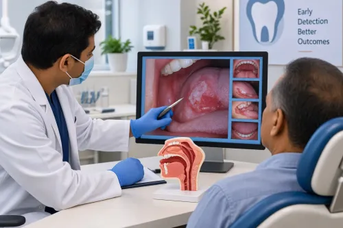
Expect the first visit to establish whether 3DCRT fits your tumour’s location, treatment goal and nearby organs. The radiation oncologist reviews scans, pathology and medical history, explains benefits and risks, and discusses the fraction schedule and possible chemotherapy.
1. Consultation: You receive an examination and treatment explanation, including expected side effects, skin care, nutrition and follow-up. Ask who will coordinate urgent problems during treatment.
2. CT simulation: You lie in the treatment position while a planning CT maps your body in three dimensions. A mould, mask or other immobilisation device helps reproduce that position; small skin marks or tattoos may guide setup. Contrast is used only when clinically needed.
3. Treatment planning: The oncologist outlines the tumour, treatment margin and organs at risk on the CT images. Physicists calculate beam angles, shaped fields and dose, then the oncologist approves the plan. Ask whether the centre documents an independent physics check.
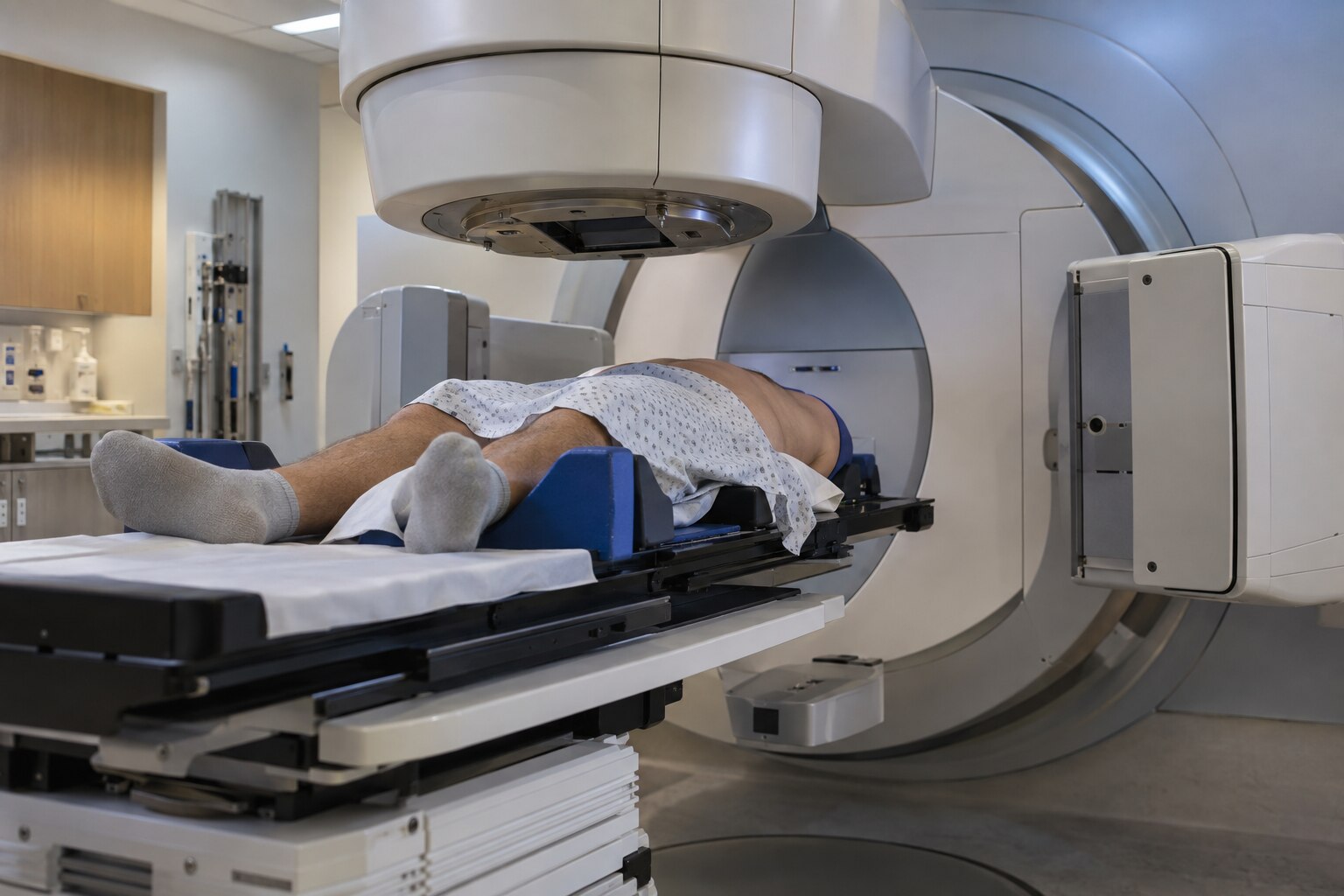
4. Daily positioning: Staff align your marks and immobilisation device on the treatment couch. They may use image-guided verification to check anatomy and adjust your position before radiation; movement or organ filling can affect accuracy.
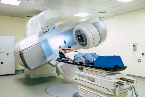
5. Radiation delivery: Each session is painless and commonly takes several minutes, though appointments last longer for setup. You remain alone in the room while the machine delivers shaped beams, with staff watching and speaking through an intercom. External-beam treatment does not make you radioactive. Most schedules use weekday fractions over several weeks.
Questions to Ask a Pune or PCMC Radiation Team
Compare the proposed plan, not the machine name. Ask what the treatment intent is—cure, recurrence-risk reduction after surgery, or symptom relief—and which organs at risk are being protected.
- Which dose will the spinal cord, kidneys, lungs, bowel, bladder or parotid glands receive?
- How will breathing or tumour motion be managed: breath-hold, respiratory gating, four-dimensional CT or image guidance?
- Would IMRT, VMAT or proton therapy materially improve normal-tissue protection for this tumour?
- Will a physicist complete an independent or documented dose check, and will image-guided verification confirm positioning before treatment?
- Who will manage skin, swallowing, bowel, urinary or fatigue problems, and whom do you call between visits?
- What follow-up schedule checks recurrence and late effects after treatment ends?
| Plan type | What to compare | When it may fit |
|---|---|---|
| 3DCRT | Shaped beams, target coverage and organ doses on the dose-volume report | A tumour with manageable geometry and motion |
| IMRT or VMAT | Whether improved conformity lowers dose to nearby organs enough to justify added complexity | A concave target or critical organ close to the tumour |
| Proton therapy | Whether the lower exit dose changes your specific plan and access burden | Selected cases after a comparative plan review |
The 3dcrt radiotherapy benefits in Pune depend on this evidence, not proximity alone; shorter travel helps, but it cannot replace a dose-plan and safety comparison. Dr Siddhesh Tryambake can be asked to explain these trade-offs in figures you can understand.
Related service
Radiation Therapy Treatment in PCMC Advanced 3DCRT Radiotherapy at Phoenix Cancer Care Clinic, PCMC At Phoenix Cancer Care Clinic, PCMC, Dr. View service → |
Frequently asked questions
How does CT-based 3DCRT target a tumour?
A planning CT creates a three-dimensional map of the tumour and nearby normal tissue. The radiation oncologist outlines these structures, selects beam directions and shapes each beam to match the tumour’s outline.
What are the benefits and limits of 3DCRT for different cancers?
3DCRT can deliver a planned dose while reducing radiation outside the tumour. Its suitability depends on the tumour’s size, shape, location and distance from sensitive organs; IMRT, VMAT or stereotactic treatment may provide better conformality in some cases.
When is 3DCRT preferred over other radiation techniques?
A radiation oncologist compares 3DCRT with IMRT, VMAT and stereotactic radiotherapy using the tumour location, treatment intent, dose distribution and nearby organs at risk.
What happens before and during a 3DCRT treatment course?
You have a planning CT, receive positioning instructions and may receive skin marks or an immobilisation device. The team then completes treatment planning before delivering scheduled radiation sessions in the treatment position.
What should I ask a Pune or PCMC radiation team about 3DCRT?
Ask why 3DCRT suits your cancer, which organs require protection, what alternatives were compared, how many sessions are planned, what side effects to report and how follow-up will work.
Keywords