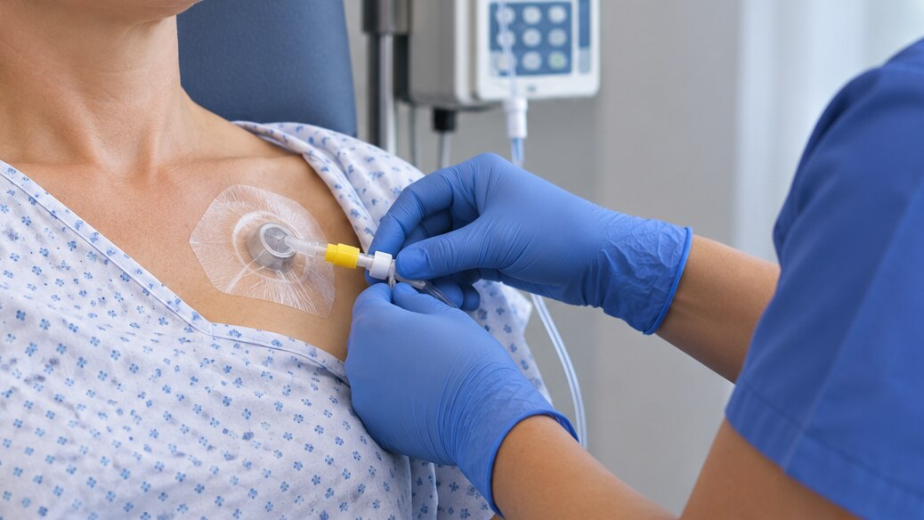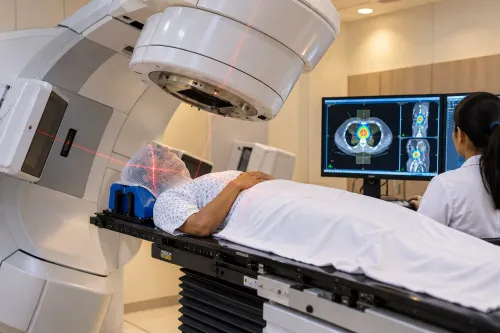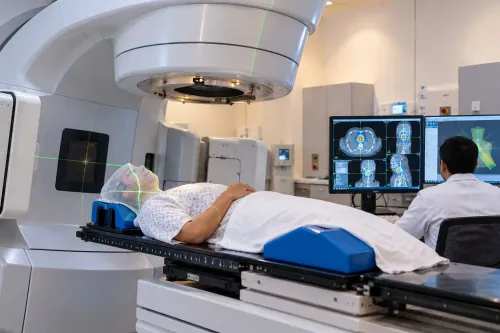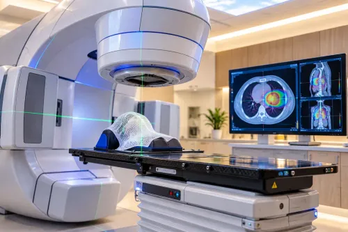
An IGRT visit includes more than time under the treatment machine: the team confirms your identity and treatment site, reproduces your planned position, takes verification images and adjusts alignment before radiation is delivered. By the end, you will know what happens from registration through follow-up, how planning and imaging fit together, and which questions and records to bring to your appointment.
Key takeaways
- Allow 60–90 minutes for registration, positioning, imaging and treatment.
- Bring scans, pathology reports, medication lists and previous radiation records.
- IGRT uses images to verify position; IMRT and VMAT describe beam delivery.
- Ask how breathing motion, bladder filling and body position will be managed.
What happens during an image-guided radiotherapy appointment?
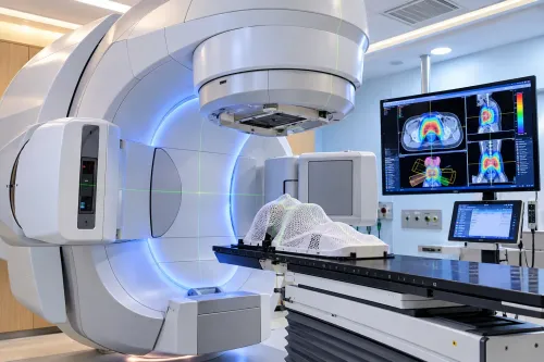
Allow 60–90 minutes for an image guided radiotherapy appointment in PCMC, although the radiation beam may run for only a few minutes. Extra time goes into registration, positioning and image matching.
- Check in and confirm your identity, treatment site and scheduled fraction with the radiation therapy team.
- Change into the required clothing and remove metal items if instructed. Tell staff about new pain, breathing difficulty, pregnancy, or trouble holding the required position.
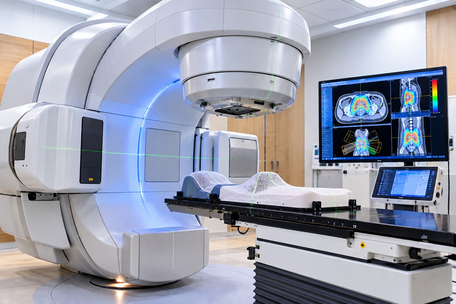
- Lie on the treatment couch in the same position used during simulation. Staff may fit a mask, vacuum cushion, body mould, or other immobilisation device.
- The machine takes verification images, such as kV X-rays, portal images, or cone-beam CT. These checks compare your anatomy, markers, or implanted fiducials with the approved treatment plan.
- Remain still while the team reviews the images and adjusts the couch or your position. Staff may repeat imaging when the target is not aligned.
- The team leaves the room, monitors you through cameras and an intercom, and delivers radiation. You do not feel the radiation, but the machine may rotate and make sounds.
- Staff may take another image during treatment when movement monitoring is part of the plan.
After delivery, the team checks how you feel, removes immobilisation equipment, and gives instructions for the next fraction. You can usually leave immediately; IGRT does not make you radioactive. Confirm the next appointment and whom to call if symptoms develop or you cannot maintain the treatment position.
How is radiation therapy planned before the first treatment?
Before the first fraction, the team turns your prescription into a reproducible treatment plan. Radiation therapy planning in PCMC usually starts with a simulation CT while you lie in the same position used on the treatment couch, often with an immobilisation mask, vacuum cushion, knee support or body mould.
| Scan | Planning purpose |
|---|---|
| Simulation CT | Shows anatomy in treatment position and provides the measurements used for dose calculation. |
| Diagnostic CT or MRI | Adds detail for tumour definition and is registered with the simulation CT when appropriate. |
| PET/CT | Highlights metabolically active disease when it is relevant to target definition. |
Your position is recorded with skin marks, couch coordinates and instructions such as breath-holding, controlled breathing or bladder preparation. The radiation oncologist then contours the visible tumour, clinical target volume and planning target volume, plus organs at risk such as the spinal cord, lungs, heart, kidneys or bowel.
A dosimetrist or medical physicist calculates beam angles, energy, intensity, monitor units and dose distribution. The plan is reviewed against target coverage and normal-tissue limits; a tighter target margin does not justify exceeding an organ constraint. For radiation therapy planning PCMC teams also review image guidance requirements, including the images needed before treatment.
Before approval, the team checks the prescription, patient identity, treatment site, contours, dose-volume results and plan parameters. An independent or secondary dose calculation follows, and machine-specific quality assurance tests whether an IMRT, VMAT or stereotactic plan can be delivered accurately.
Which images are taken, and how does IGRT differ from IMRT or VMAT?
A verification image answers a positioning question before radiation is delivered: is the patient, treatment site, and planned radiation geometry aligned closely enough to treat safely?
- kV X-rays show bones and implanted markers, confirming couch alignment and the target’s position against the planned reference.
- MV portal images check the treatment field, beam shape, and machine-to-patient geometry; they provide less soft-tissue detail.
- Cone-beam CT shows three-dimensional anatomy, confirming the tumour region, nearby organs, and changes in filling or swelling.
- Surface imaging checks the patient’s external contour and posture without additional radiation, but it cannot reliably show an internal tumour.
- Ultrasound or fiducial-marker imaging confirms target location when those tools suit the treatment site.
The team matches the new image with the simulation image and applies a couch correction when needed. Imaging can occur before each fraction and during treatment for selected plans.
| Feature | IGRT | IMRT | VMAT |
|---|---|---|---|
| Purpose | Verifies position and target location | Shapes dose with different beam intensities | Shapes dose while the gantry rotates |
| What changes | Patient or couch alignment | Multileaf collimator pattern and intensity | Intensity, leaf positions, dose rate, and gantry position |
| Can they combine? | Yes | Yes | Yes |
IGRT is therefore a verification method, not a beam-delivery technique. A course using 3D conformal radiation therapy, IMRT, VMAT, or stereotactic body radiation therapy can all include image guidance. Availability of MRI guidance, fiducial tracking, or motion imaging requires confirmation from the clinic.
How are imaging and motion management matched to the cancer site?
The cancer site determines how you are positioned, how often images are taken, and whether you must control your breathing. If you are comparing image guided cancer treatment pcmc options, ask whether the planned checks match the movement of your tumour and nearby organs.
- Lung: You may lie with arms raised while cone-beam CT or kV images verify the chest position. Breath-hold, shallow breathing, or four-dimensional CT planning may be used because the tumour moves as you breathe.
- Liver: Treatment may use breath-hold, respiratory gating, or repeated imaging to track movement. A fiducial marker or implanted reference marker is needed only when prescribed.
- Prostate: A consistent bladder and rectum condition helps reproduce the planned anatomy. Daily imaging is common because prostate position changes with bladder filling and rectal gas.
- Breast: You may lie supine with the arm above your head. Deep-inspiration breath-hold can move the heart away from the breast, especially for left-sided treatment.
- Head and neck: A thermoplastic mask keeps your head and shoulders still. Daily image matching checks this close fit because small shifts can affect the brain, throat, spinal cord, or salivary glands.
- Abdominal sites: Fasting, bladder filling, breath control, or image checks before each fraction can reduce organ movement and day-to-day variation.
Ask the team which instruction applies to you, how long you must hold your breath, and what happens if you cannot maintain it.
What should you confirm and bring to a PCMC radiation oncology clinic?
Before choosing an image guided radiation therapy clinic in PCMC, confirm exactly what will happen, what you must bring, and who manages problems between visits.
Bring these records in paper or digital form:
- Government photo ID, referral letter, biopsy and pathology reports
- CT, MRI or PET-CT reports and image files on a disc or USB drive
- Previous radiation summaries, chemotherapy details, medication list and allergy history
- Insurance documents and contact details for your referring doctor
Ask whether you need fasting, a full or empty bladder, bowel preparation, contrast, or implanted fiducial markers. Tell the team about diabetes, kidney disease, contrast reactions, pregnancy possibility, pain, or difficulty lying still. Confirm the expected number of fractions, visit frequency, appointment duration, and whether an immobilisation mask or vacuum cushion will be made.
Ask what imaging and motion-control equipment is actually available:
| Option | What it checks | Question to ask |
|---|---|---|
| kV or MV X-rays | Bone or marker position | Is imaging taken before every fraction? |
| Cone-beam CT | Three-dimensional anatomy and organ filling | Is it performed on the treatment machine? |
| Surface imaging or respiratory monitoring | External position or breathing motion | Will you receive instructions to hold or control your breath? |
An IGRT label does not prove that MRI guidance, SBRT, brachytherapy, PET-CT or motion management is available. Dr Siddhesh Tryambake can clarify which technique fits your prescription and what the clinic provides on-site.
Before attending a radiation oncology clinic Pimpri-Chinchwad, obtain a named contact for missed appointments, new symptoms, fever, uncontrolled pain, or inability to maintain the required position. Confirm on-treatment review dates and how follow-up scans and reports will be shared.
Related service
Radiation Therapy Image Guiding Radiation Therapy Image-Guided Radiation Therapy (IGRT) in PCMC Advanced Image-Guided Radiation Therapy at Phoenix Cancer Care Clinic, PCMC At Phoenix Cancer Care... View service → |
Frequently asked questions
What happens during an image-guided radiotherapy appointment?
You complete registration, change if required, lie on the treatment couch, and receive positioning checks. The team takes verification images, compares them with your planning scans, makes approved adjustments, and delivers radiation. Allow 60–90 minutes even when the beam runs for only a few minutes.
How is radiation therapy planned before the first treatment?
The team reviews your diagnosis and scans, performs a planning CT in the treatment position, defines the tumour and nearby organs, and creates a dose plan. You may receive positioning marks or an immobilisation device. The plan is checked before treatment begins.
Which images are used during IGRT, and how does it differ from IMRT or VMAT?
IGRT uses treatment-day images, such as cone-beam CT or paired X-rays, to verify anatomy and position. IMRT shapes radiation intensity across beam fields, while VMAT delivers changing beam angles as the machine rotates. IGRT concerns image verification; IMRT and VMAT concern dose delivery.
How are imaging and motion management matched to the cancer site?
The team selects imaging and positioning methods based on the treatment area. Abdominal and chest treatments may require breath-hold, respiratory monitoring or daily soft-tissue imaging. Pelvic treatments depend on consistent bladder and bowel preparation. Head, neck and spine treatments require precise immobilisation.
What should you confirm and bring to a PCMC radiation oncology clinic?
Confirm the appointment time, preparation instructions, contrast requirements, clothing guidance and whether you need a full bladder or empty rectum. Bring identification, referral documents, pathology reports, scan discs and reports, medication details, allergy information, and previous treatment records.
Keywords